
Histology Lab
Elena Aikawa
Members
- Elena Aikawa
- Alexandria Biondo
Lab Motto
Preserving the Image
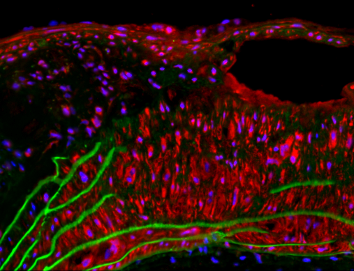
Immunofluorescence staining for Cathepsin S
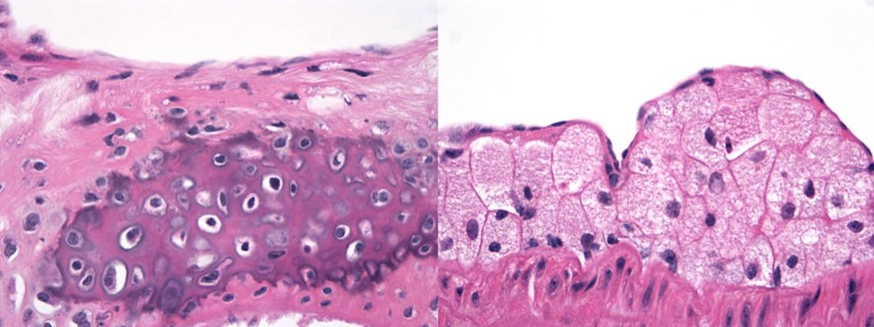
Haematoxylin and eosin (H&E) staining of mouse atherosclerotic plaques. Left, an advanced plaque with calcification. Right, Foam cell accumulation in an early stage plaque.
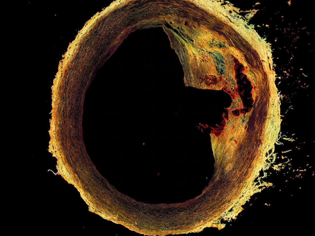
Alizarin red staining of human lower extremity artery.
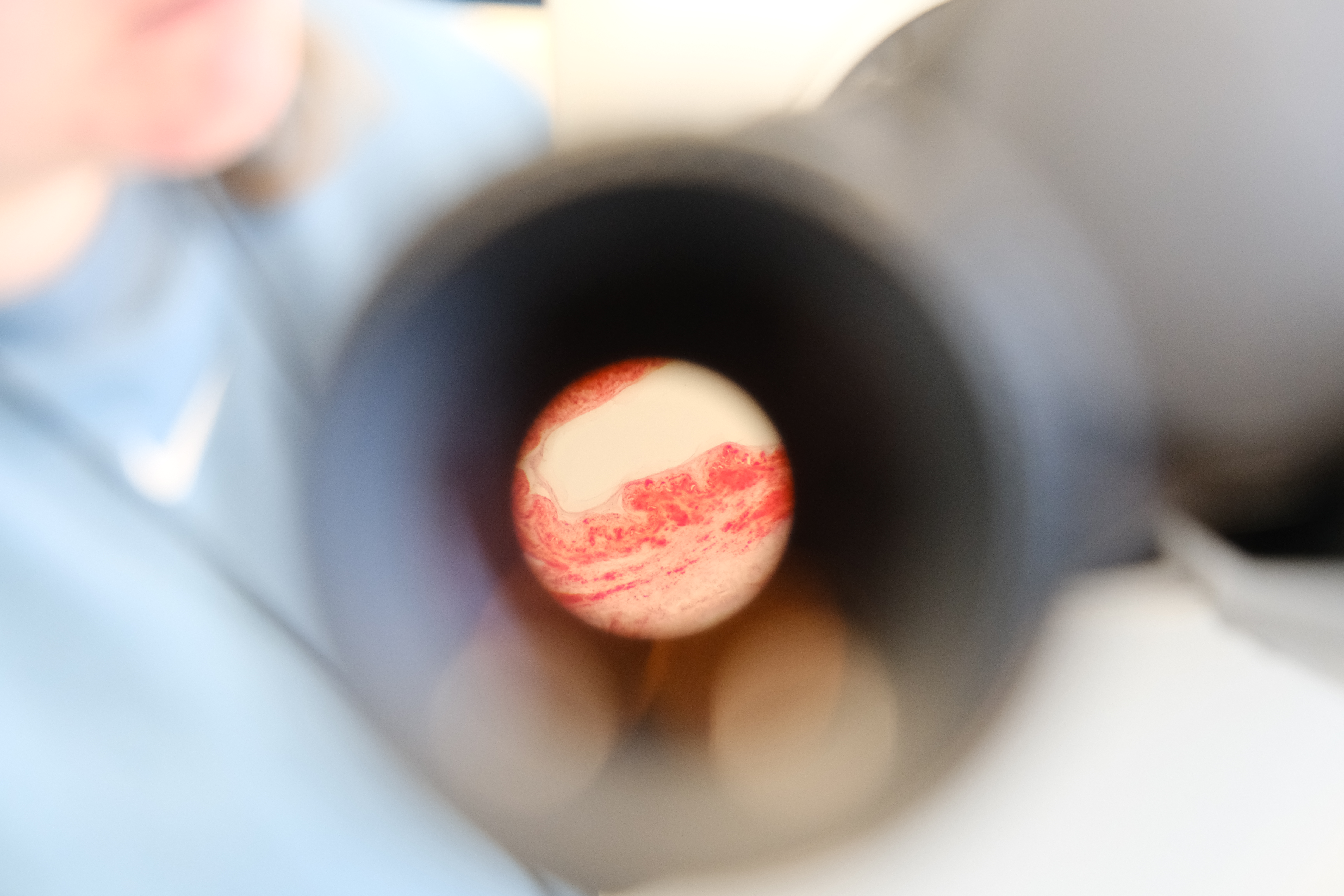
Picrosirius Red Stain of a carotid artery cross-section
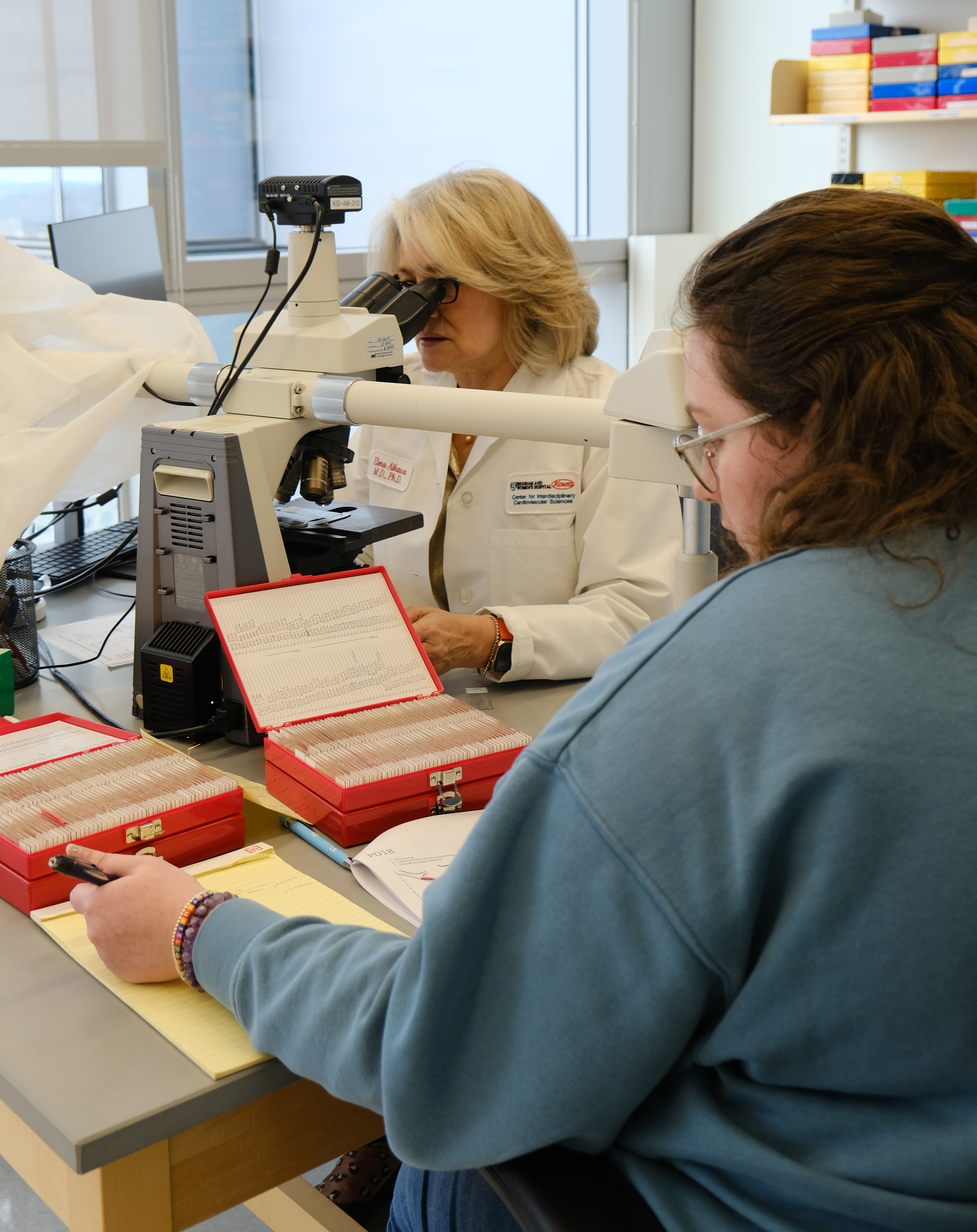
Dr. Elena Aikawa and histology technician Alexandria (Lexi) Biondo examining slides together.
About Us
Our lab is devoted to excellence in the preservation and study of the microscopic anatomy of cells and tissues. Involved with each step of the histological process, we preserve, cut, and save tissue samples. Using a variety of staining techniques from H&E to immunohistochemistry and immunofluorescence we accurately highlight specific types of tissue, proteins or sections. With access to other high powered microscopes including polarizing, fluorescent, and confocal microscopes we quantify the tissues and cells to obtain precise and accurate data to complement and contribute to the research being conducted at CICS. Using our Omnyx VL4 slide scanner, we can capture high definition images of whole tissue sections on slides to further our quantification and analysis. Our laser microdissection microscope provides the opportunity for us to further our experimental assistance by precisely cutting and sectioning small portions of tissues to collect an aggregate of a specific tissue type. We can further detect and analyze nanoparticles within tissues by employing our high-powered instruments. We pride ourselves on our techniques, on our skill in developing defined and complete stains, and our meticulous attention to detail.